Video
In vitro motility assay
Consists in observing fluorescently labeled actin filaments as they get propelled by myosin molecules adhered randomly to a nitrocellulose-coated coverslip. The velocity of actin filament propulsion is determined in presence of MgATP. (TRITC-labeled skeletal muscle actin filaments propelled by smooth muscle myosin).
Fluorescently labeled actin filaments propelled by smooth muscle myosin purified from chicken gizzard.
Laser trap: unbinding assay

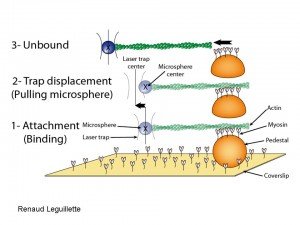
A single beam laser trap assay is used to capture a polystyrene bead (blue) to which a TRITC-labeled actin filament isttached. 1: The actin is brought in contact with unphosphorylated myosin that randomly coats a pedestal on a coverslip. 2: The pedestal/coverslip is then moved away from the laser trap at a constant and slow velocity, thereby dragging the trapped bead away from the trap center as a result of the attachment of unphosphorylated myosin to actin. 3: When the force exerted by the trap on the bead is greater than the force of binding of unphosphorylated myosin to actin, the bead snaps back into the trap, its unloaded position. The unbinding force (Funb) is then calculated as the maximal distance between the bead and the trap center (max Δd) multiplied by the trap stiffness calculated by the Stokes force method. (see video)

